No items found.

Please help support Ninja Nerd to continue creating free medical videos. All donations we receive are put directly into content creation so we are able to continue providing these videos for free. Thanks!

Ninja Nerds!
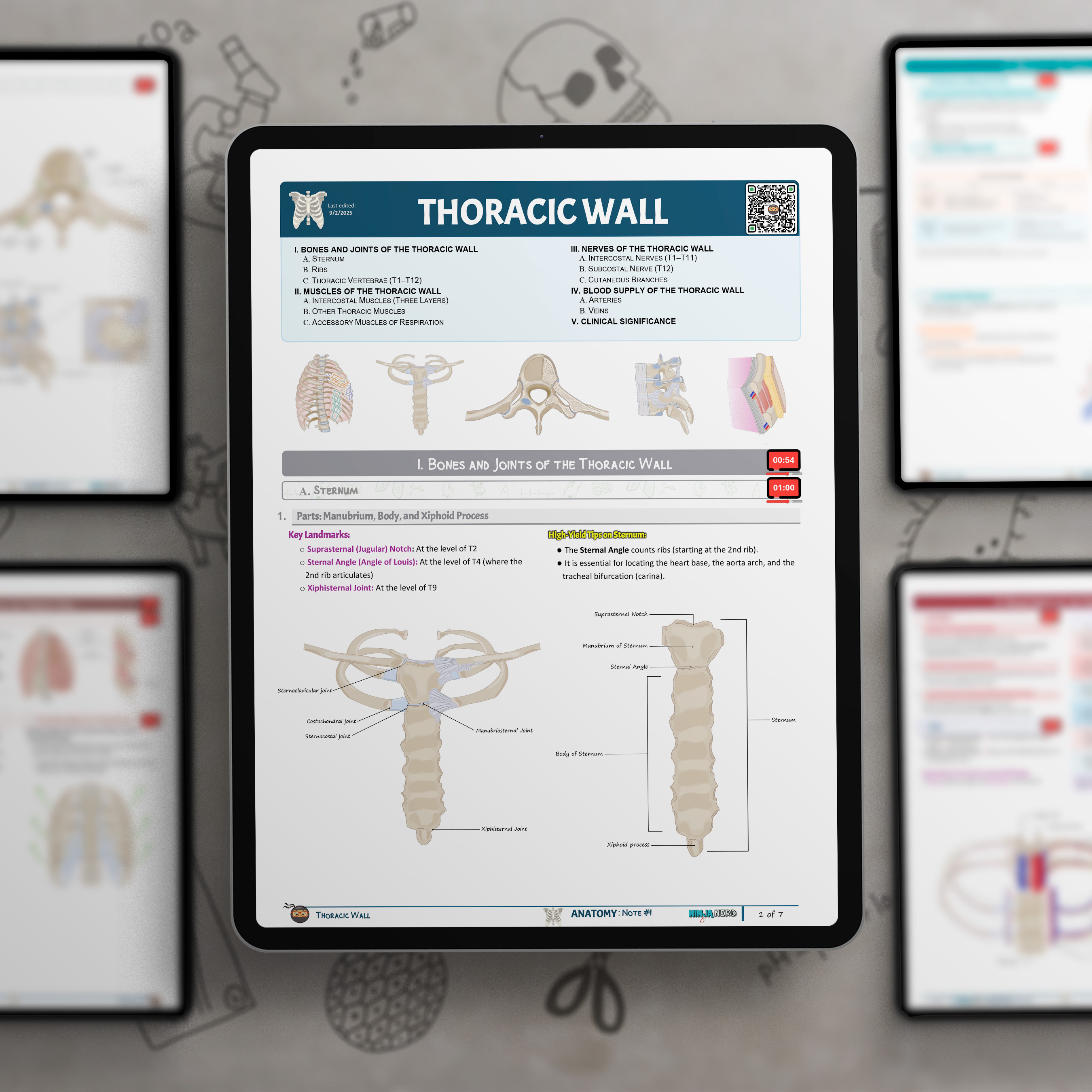
In this lecture, Professor Zach Murphy presents on the anatomy of the Thoracic Wall. The session begins with a detailed digital presentation using professional anatomical illustrations to explore the key components of the thoracic wall, including the sternum, ribs, thoracic vertebrae, and associated joints such as the costovertebral, costotransverse, and sternocostal joints. Clinical landmarks like the sternal angle (Angle of Louis) and xiphisternal joint are emphasized for their anatomical and procedural relevance.
We then cover the muscles of the thoracic wall, including the external, internal, and innermost intercostals, along with supporting structures like the transversus thoracis, subcostal muscles, and accessory muscles of respiration. Their roles in breathing mechanics and respiratory compensation are clearly explained.
The lecture discusses the neurovascular structures of the thoracic wall, focusing on the intercostal nerves, internal thoracic artery, posterior intercostal vessels, and the azygos and hemiazygos venous systems. The presentation highlights significant clinical correlations such as intercostal nerve blocks, costochondritis, rib fractures, flail chest, and safe zones for chest tube placement.
To conclude, we transition to @Anatomage where we examine the same thoracic wall anatomy on real digital cadaveric images. This final portion of the lecture brings clinical context and three-dimensional realism to reinforce the anatomical relationships covered in the digital illustrations.
Sorry, but there are currently no notes available for this video.
Check back again soon, or contact us to find out when they will be available.